
Larynx
Below are microphotographs taken in the practical histology laboratory of the University of Sonora.
DOI:
https://doi.org/10.59420/remus.13.2025.277Keywords:
Larynx , Histology, Vocal cordsAbstract
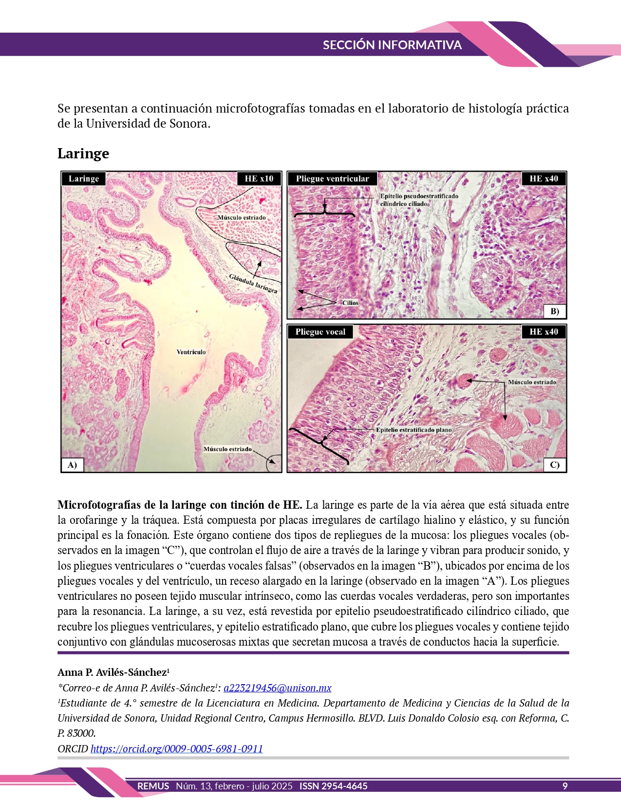
Microphotographs of the larynx with HE staining. The larynx is part of the airway located between the oropharynx and the trachea. It is composed of irregular plates of hyaline and elastic cartilage, and its main function is phonation. This organ contains two types of mucosal folds: the vocal folds (seen in image "C"), which control airflow through the larynx and vibrate to produce sound, and the ventricular folds or "false vocal folds" (seen in image "B"), located above the vocal folds and the ventricle, an elongated recess in the larynx (seen in image "A"). The ventricular folds do not have intrinsic muscle tissue, like the true vocal folds, but are important for resonance. The larynx, in turn, is lined by ciliated pseudostratified columnar epithelium, which lines the ventricular folds, and stratified squamous epithelium, which covers the vocal folds and contains connective tissue with mixed mucous glands that secrete mucus through ducts to the surface.
Downloads
References
Ross MH, Pawlina W. Histología: texto y atlas con biología celular y molecular. 16ª ed. Barcelona: Wolters Kluwer; 2021.
Downloads
Published
How to Cite
Issue
Section
License
Copyright (c) 2024 REMUS - Revista Estudiantil de Medicina de la Universidad de Sonora

This work is licensed under a Creative Commons Attribution-NonCommercial-NoDerivatives 4.0 International License.
