
Informative section - Lung
The information section is a section created for the publication of microscopic images with the aim of promoting a culture of research and publication beginning in preclinical semesters. The authors of these images are undergraduate medical students from the University of Sonora who are completing their laboratory work in Histology, a subject taught by Dr. Guillermo López Cervantes, with the support of the MPSS.
DOI:
https://doi.org/10.59420/remus.9.2023.153Keywords:
Respiratory bronchiolar unit, Gas exchange, Type I and II pneumocytes, Interalveolar septumAbstract
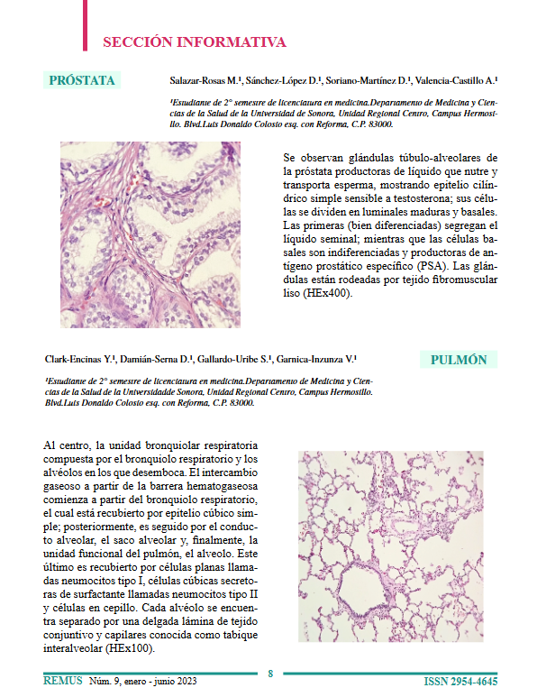
At the center, the respiratory bronchiolar unit consists of the respiratory bronchiole and the alveoli into which it empties. Gas exchange across the blood-gas barrier begins at the respiratory bronchiole, which is lined by simple cuboidal epithelium; it is then followed by the alveolar duct, the alveolar sac, and finally, the functional unit of the lung, the alveolus. The latter is lined by flat cells called type I pneumocytes, surfactant-secreting cuboidal cells called type II pneumocytes, and brush cells. Each alveolus is separated by a thin layer of connective tissue and capillaries known as the interalveolar septum (HEx100).
Downloads
References
Ross MH, Pawlina W. Histología: texto y atlas con biología celular y molecular. 16ª ed. Barcelona: Wolters Kluwer; 2021.
Downloads
Published
How to Cite
Issue
Section
License
Copyright (c) 2023 REMUS - Revista Estudiantil de Medicina de la Universidad de Sonora (Journal of Medical Students' of the University of Sonora)

This work is licensed under a Creative Commons Attribution-NonCommercial-NoDerivatives 4.0 International License.
