
Músculo estriado esquelético
DOI:
https://doi.org/10.59420/remus.10.2023.181Palabras clave:
Músculo estriado, Miofibras, Tejido conectivo, Tinción de MassonResumen
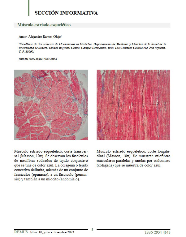
El músculo estriado esquelético presenta una organización estructural definida que puede apreciarse mediante tinción de Masson a 10x. En cortes longitudinales, se observan miofibras dispuestas de forma paralela, unidas entre sí por endomisio compuesto de colágena, que se tiñe de color azul. En cortes transversales, los fascículos musculares están delimitados por tejido conectivo también teñido de azul, que corresponde al epimisio (conjunto de fascículos), perimisio (alrededor de un fascículo) y endomisio (alrededor de un miocito). Esta disposición evidencia la complejidad anatómica y funcional del músculo esquelético.
Descargas
Citas
Ross MH, Pawlina W. Histología: texto y atlas con biología celular y molecular. 16ª ed. Barcelona: Wolters Kluwer; 2021.
Publicado
Cómo citar
Número
Sección
Licencia
Derechos de autor 2023 REMUS - Revista Estudiantil de Medicina de la Universidad de Sonora

Esta obra está bajo una licencia internacional Creative Commons Atribución-NoComercial-SinDerivadas 4.0.
