
Skeletal striated muscle
DOI:
https://doi.org/10.59420/remus.10.2023.181Keywords:
Striated muscle, Myofibers, Connective tissue, Masson stainingAbstract
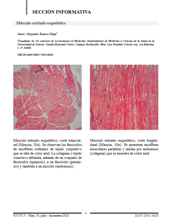
Skeletal striated muscle exhibits a well-defined structural organization, observable through Masson staining at 10x magnification. In longitudinal sections, parallel muscle fibers are seen, joined by endomysium composed of collagen, stained blue. In transverse sections, muscle fascicles are surrounded by blue-stained connective tissue, which corresponds to the epimysium (surrounding groups of fascicles), perimysium (around a single fascicle), and endomysium (around individual myocytes). This arrangement highlights the anatomical and functional complexity of skeletal muscle tissue.
Downloads
References
Ross MH, Pawlina W. Histología: texto y atlas con biología celular y molecular. 16ª ed. Barcelona: Wolters Kluwer; 2021.
Downloads
Published
How to Cite
Issue
Section
License
Copyright (c) 2023 REMUS - Revista Estudiantil de Medicina de la Universidad de Sonora (Journal of Medical Students' of the University of Sonora)

This work is licensed under a Creative Commons Attribution-NonCommercial-NoDerivatives 4.0 International License.
